- Hip Basics
- Hip Injuries & Conditions
- Non-surgical & Regenerative Medicine
- Surgeries Performed by Dr. Milligan
- Surgical Videos
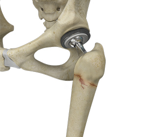
Hip Joint
The hip joint is the largest weight-bearing joint in the human body. It is also referred to as a ball and socket joint and is surrounded by muscles, ligaments, and tendons. The thigh bone or femur and the pelvis join to form the hip joint.
Any injury or disease of the hip will adversely affect the joint's range of motion and ability to bear weight.
The hip joint is made up of the following:
- Bones and joints
- Ligaments of the joint capsule
- Muscles and tendons
- Nerves and blood vessels that supply the bones and muscles of the hip
Bones and Joints
The hip joint is the junction where the hip joins the leg to the trunk of the body. It is comprised of two bones: the thigh bone or femur and the pelvis which is made up of three bones called ilium, ischium, and pubis. The ball of the hip joint is made by the femoral head while the socket is formed by the acetabulum. The Acetabulum is a deep, circular socket formed on the outer edge of the pelvis by the union of three bones: ilium, ischium, and pubis. The lower part of the ilium is attached by the pubis while the ischium is considerably behind the pubis. The stability of the hip is provided by the joint capsule or acetabulum and the muscles and ligaments which surround and support the hip joint.
The head of the femur rotates and glides within the acetabulum. A fibrocartilagenous lining called the labrum is attached to the acetabulum and further increases the depth of the socket.
The femur or thigh bone is one of the longest bones in the human body. The upper part of the thigh bone consists of the femoral head, femoral neck, and greater and lesser trochanters. The head of the femur joins the pelvis (acetabulum) to form the hip joint. Next, to the femoral neck, there are two protrusions known as greater and lesser trochanters which serve as sites of muscle attachment.
Articular cartilage is the thin, tough, flexible, and slippery surface lubricated by synovial fluid that covers the weight-bearing bones of the body. It enables smooth movements of the bones and reduces friction.
Ligaments
Ligaments are fibrous structures that connect bones to other bones. The hip joint is encircled with ligaments to provide stability to the hip by forming a dense and fibrous structure around the joint capsule. The ligaments adjoining the hip joint include:
- Iliofemoral ligament: This is a Y-shaped ligament that connects the pelvis to the femoral head at the front of the joint. It helps in limiting the over-extension of the hip.
- Pubofemoral ligament: This is a triangular shaped ligament that extends between the upper portion of the pubis and the iliofemoral ligament. It attaches the pubis to the femoral head.
- Ischiofemoral ligament: This is a group of strong fibers that arise from the ischium behind the acetabulum and merge with the fibers of the joint capsule.
- Ligamentum teres: This is a small ligament that extends from the tip of the femoral head to the acetabulum. Although it has no role in hip movement, it does have a small artery within that supplies blood to a part of the femoral head.
- Acetabular labrum: The labrum is a fibrous cartilage ring which lines the acetabular socket. It deepens the cavity, increasing the stability and strength of the hip joint.
Muscles and Tendons
A long tendon called the iliotibial band runs along the femur from the hip to the knee and serves as an attachment site for several hip muscles including the following:
- Gluteals: These are the muscles that form the buttocks. There are three muscles (gluteus minimus, gluteus maximus, and gluteus medius) that attach to the back of the pelvis and insert into the greater trochanter of the femur.
- Adductors: These muscles are located in the thigh which helps in adduction, the action of pulling the leg back towards the midline.
- Iliopsoas: This muscle is located in front of the hip joint and provides flexion. It is a deep muscle that originates from the lower back and pelvis and extends up to the inside surface of the upper part of the femur.
- Rectus femoris: This is the largest band of muscles located in front of the thigh. They also are hip flexors.
- Hamstring muscles: These begin at the bottom of the pelvis and run down the back of the thigh. Because they cross the back of the hip joint, they help in extension of the hip by pulling it backward.
Nerves and Arteries
Nerves of the hip transfer signals from the brain to the muscles to aid in hip movement. They also carry the sensory signals such as touch, pain, and temperature back to the brain.
The main nerves in the hip region include the femoral nerve in the front of the femur and the sciatic nerve at the back. The hip is also supplied by a smaller nerve known as the obturator nerve.
In addition to these nerves, there are blood vessels that supply blood to the lower limbs. The femoral artery, one of the largest arteries in the body, arises deep in the pelvis and can be felt in front of the upper thigh.
Hip Movements
All of the anatomical parts of the hip work together to enable various hip movements. Hip movements include flexion, extension, abduction, adduction, circumduction, and hip rotation.

Femoroacetabular Impingement
Femoroacetabular impingement (FAI) is a condition characterized by excessive friction in the hip joint from the presence of bony irregularities. These cause pain and decreased range of hip motion.

Ischiofemoral Impingement
Ischiofemoral impingement is a condition in which there is an abnormal contact (impingement) between the soft tissues of the thigh bone and hip due to the narrowing of space between the lesser trochanter and ischium, resulting in significant hip pain.

Hip Ligament Injuries
Injuries to the hip ligaments are commonly called a hip sprain and can range from minor tears of the ligaments to more serious injuries involving the hip muscles, tendons or bone.

Femoral Subchondral Cysts
Femoral subchondral cysts are fluid-filled sacs or spaces that form in the femur (thighbone) side of the hip joint. Subchondral refers to the layer of bone just below the cartilage in a joint.

Borderline Hip Dysplasia
Hip dysplasia is a medical condition where the acetabulum (hip socket) does not fully cover the ball-like head at the top of the femur (thighbone). Most people who have hip dysplasia are born with it.
Periprosthetic Hip Fractures
Hip replacement is a surgical procedure in which the damaged cartilage and bone are removed from the hip joint and replaced with artificial components. Any resulting fractures or breaks in the bone around the implant are called periprosthetic hip fractures.

Snapping Hip Syndrome
Snapping hip syndrome is a condition in which you hear or feel a snapping sound in the hip when you swing your legs, run, walk or get up from a chair. The sound can be experienced in the back, front or side of the hip.

Hip Bursitis
Hip bursitis is a painful condition caused by the inflammation of a bursa in the hip. Bursae are fluid-filled sacs present in the joints between bone and soft tissue to reduce friction and provide cushioning during movement.

Avascular Necrosis
Avascular necrosis, also called osteonecrosis, is a condition in which bone death occurs because of inadequate blood supply to it. Lack of blood flow may occur when there is a fracture in the bone or a joint dislocation that may damage nearby blood vessels.
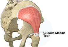
Gluteus Medius Tear
A gluteus medius tear is the partial or complete rupture of the gluteus medius muscle due to severe muscle strain. Gluteus medius tears often occur at the tendinous attachment to the greater trochanter of the femur bone.

Hip Labral Tear
A hip labral tear is an injury to the labrum, the cartilage that surrounds the outside rim of your hip joint socket.

Chondral Lesions or Injuries of the Hip
Chondral injuries can result from various hip conditions such as labral tears, loose bodies, posterior dislocation, slipped capital femoral epiphysis (SCFE), dysplasia, osteonecrosis, and degenerative arthritis.

Hip Instability
Injury or damage to these structures can lead to a condition called hip instability when the joint becomes unstable.

Hip Abductor Tears
Hip abductors are a major group of muscles found in the buttocks. It includes the gluteus maximus, gluteus medius, gluteus minimus, and tensor fascia lata muscles.

Developmental Dysplasia
Developmental dysplasia of the hip (DDH) or hip dysplasia is a condition that is seen in infants and young children because of developmental problems in the hip joint. The femur (thighbone) partially or completely slips out of the hip socket leading to dislocation at the hip joint.

Slipped Capital Femoral Epiphysis
Slipped capital femoral epiphysis (SCFE) is an unusual disorder of the hip where the ball at the upper end of the thighbone (femur) slips in a backward direction. This is caused due to weakness of the growth plate. This condition is commonly caused during accelerated growth periods such as the onset of puberty.

Legg-Calve-Perthes Disease
Legg-Calve-Perthes disease (LCPD) or Perthes disease is a disorder of the hip that affects children, usually between the ages of 4 and 10. It usually involves one hip, although it can occur on both sides in some children. It occurs more commonly in boys than girls.

Osteoarthritis of the Hip
Osteoarthritis, also called degenerative joint disease, is the most common form of arthritis. It occurs most often in the elderly. This disease affects the tissue covering the ends of bones in a joint called cartilage.

Groin Injuries in Athletes
Groin injuries are injuries sustained by athletes during sports activity. Groin injuries comprise about 2 to 5 percent of all sports injuries. The most common kind of groin injury is a groin strain or a pulled groin muscle.

Hip Retroversion
Hip retroversion also known as femoral retroversion is a rotational deformity in which the hip is rotated outward in relation to the knee. Hip retroversion can develop in one or both legs in both children and adults.

Periprosthetic Hip Infection
A very small percentage of patients (less than 1%) who undergo hip replacement may develop an infection around the hip joint following surgery. This infection is called a periprosthetic hip infection.
Platelet-Rich Plasma (PRP) Injection
What is Platelet-Rich Plasma (PRP)?
Our blood consists of a liquid component known as plasma. It also consists of three main solid components which include the red blood cells (RBCs), white blood cells (WBCs), and platelets. Platelets play an important role in forming blood clots. They also consist of special proteins, known as growth factors, which help with our body’s healing process. Platelet-rich plasma or PRP is a high concentration of platelets and plasma. A normal blood specimen contains only 6% platelets, while platelet-rich plasma contains 94% of platelets and 5 to 10 times the concentration of growth factors found in normal blood, thus greater healing properties.
What are the Indications for PRP Injections?
PRP is a relatively new method of treatment for several orthopedic conditions such as muscle, ligament, and tendon injuries; arthritis; and fractures. PRP injections can help alleviate painful symptoms, promote healing, and delay joint replacement surgeries.
Platelet-Rich Plasma Injection Procedure
Your doctor will first draw about 10 cc’s of blood from the large vein in your elbow. The blood is then spun in a centrifuge machine for about 10 to 15 minutes to separate the platelets from the remaining blood components. The injured part of your body is then anesthetized with a local anesthetic. The platelet-rich portion of your blood is then injected into your affected area. In some cases, your doctor may use ultrasound guidance for proper needle placement.
Post-Procedure Care following PRP Injections
It is normal to feel some discomfort at the injection site for a few days after your procedure.
- You may use cold compresses to alleviate your symptoms.
- You will be instructed to stop any anti-inflammatory medications.
- You may resume your normal activities but should avoid any strenuous activities such as heavy lifting or exercises.
Risks and Complications of PRP Injections
There are very minimal risks associated with PRP injections. Some of the potential risks include:
- Increased pain at the injection site
- Infection
- Damage to adjacent nerves or tissues
- Formation of scar tissue
- Calcification at the injection site
Ultrasound Guided Injections
Introduction
Ultrasound is a common imaging technique that employs high-frequency sound waves to create images of the organs and other internal structures of the body. These images provide the doctor valuable information which assists in diagnosis and treatment of a wide range of tendon, muscle, and joint disorders affecting the body. It is also an excellent tool for guiding the placement of needles for both diagnostic as well as therapeutic purposes. Injection of a pain medication in combination with a local anesthetic directly to the site of injury helps relieve pain. Ultrasound-guided injection provides improved accuracy for injection site location.
Advantages
The advantages of ultrasound imaging compared to other imaging techniques includes the following:
- No patient exposure to ionizing radiation
- Able to assess tendons, ligaments, and muscles under high resolution
- Provides direct visualization of the area being treated
- Ensures accurate placement of the needle to targeted areas
Indications
The indications for diagnostic ultrasound imaging technique include the following:
- Diagnose conditions such as tendon/ligament tears, inflamed bursa, compressed nerves, joint fluid, and cysts
- Assess painful pops and snaps that occur during movement
- Deliver diagnostic injections to specific targets including joints and tendon sheaths or around nerves
- Help guide needle placement during needle aspirations or injections for patients with challenging anatomical variations or people taking blood-thinning medications
- Aspiration of a ganglion cyst
- Injection into a tendon sheath or a bursa
- Administer a nerve block (diagnostic or therapeutic)
- Guide needles in percutaneous therapy for the treatment of calcific tendonitis
Procedure
During an ultrasound-guided injection, you will be asked to lie or sit down on a table depending on the injection location. A clear water-based conducting gel is applied to your skin to assist the transmission of sound waves. Your doctor moves a hand-held probe, called a transducer, over the target area. The doctor then inserts the needle into the skin under ultrasound guidance to the specified location. The transducer emits sound waves and detects the rebound echoes from the tissue. Images are created from these sound waves which are viewed on the video display screen attached to the scanner.
Risks
Ultrasound-guided injection is a relatively safe and painless procedure. Some of the associated complications include bleeding at the site of insertion and injury to adjacent structures. You can resume your normal activities immediately after the procedure.
Summary
Ultrasound-guided injection is a safe and effective technique to diagnose and treat various musculoskeletal pain conditions such as tendonitis, bursitis, and neuritis and to perform cyst aspiration. It provides high-resolution images that enhance the accuracy of needle placement without damaging the surrounding tissues.

Muscle Sparing Anterior Hip Replacement
Muscle sparing anterior hip replacement is a minimally invasive hip surgery to replace the hip joint without cutting through any muscles or tendons as compared to traditional hip replacement that involves cutting major muscles to access the hip joint.

Revision Hip Surgery
Revision hip surgery is a repeat hip surgery performed in certain patients to correct the problems or complications of previous hip surgery and overcome its limitations.

Hip Arthroscopy
Hip arthroscopy, also referred to as keyhole or minimally invasive surgery, is a procedure in which an arthroscope is inserted into your hip joint to check for any damage and repair it simultaneously.

Hip Resurfacing
The hip joint is also known as a ball and socket joint, where the ball (femoral head) of the thigh bone fits into the socket (acetabulum) of the pelvic bone.

Periacetabular Osteotomy
Periacetabular osteotomy is a surgical procedure to treat a congenital hip condition called hip dysplasia. Hip dysplasia is either present from birth or develops in the first few months of life. Patients suffering from this condition have a shallow socket (acetabulum) of the hip joint.